


Diagnostic Ultrasound for Foot Pain
Heel pain that has lingered for months, swelling around the ankle that comes and goes, or a sharp pain in the forefoot that flares on longer walks can all sound straightforward at first. In practice, foot pain is often less simple than it appears. Diagnostic ultrasound for foot pain can help clarify what is actually happening inside the soft tissues, so treatment is based on findings rather than guesswork.
For many patients, the main frustration is not just pain itself. It is uncertainty. They may already have tried rest, different shoes, online exercises or insoles without being sure whether the problem is plantar fasciitis, a tendon injury, bursitis, nerve irritation or something else entirely. A structured assessment, with ultrasound used when clinically appropriate, can make the next step much clearer.
What diagnostic ultrasound can show
Ultrasound is a real-time imaging method that is especially useful for looking at soft tissue structures in the foot and ankle. That includes tendons, ligaments, fascia, joint lining, bursae and some nerve-related problems. It can also help identify fluid, thickening, tears, inflammation and changes in tissue quality.
One of its practical advantages is that the scan can be performed dynamically. In other words, the clinician can assess the area while you move the foot, point to the painful spot, or reproduce the movement that causes symptoms. That matters because some problems are most obvious when the tissue is under load or when structures move against each other.
For foot pain, ultrasound is commonly helpful in cases such as plantar fascia pain at the heel, Achilles tendon problems, peroneal tendon irritation on the outer ankle, tibialis posterior tendon dysfunction on the inner ankle, Morton’s neuroma, bursitis, ganglion cysts and certain ligament injuries. It may also support decisions around injection therapy or rehabilitation planning.
When diagnostic ultrasound for foot pain is most useful
Not every painful foot needs imaging. A good clinical history and physical examination still come first. In many cases, an experienced podiatrist can identify the likely diagnosis through assessment alone. The value of ultrasound is that it adds detail when the findings need to be confirmed, when symptoms are persistent, or when treatment choices depend on knowing the exact structure involved.
This is particularly helpful when symptoms overlap. Heel pain, for example, is not always plantar fasciitis. Pain under the heel may come from the plantar fascia, but it can also relate to fat pad irritation, nerve involvement or nearby soft tissue inflammation. Pain around the ankle may feel like a simple strain, yet the underlying issue could be tendon degeneration, a split tear or a sheath inflammation that changes the treatment plan.
Ultrasound is also useful when a patient has had recurring symptoms despite previous treatment. If pain keeps returning, it is reasonable to ask whether the original diagnosis was complete, whether more than one structure is involved, or whether tissue changes are more advanced than expected.
What happens during assessment and scan
At South London Foot Clinic, ultrasound is not treated as a separate add-on for its own sake. It is used within a broader consult, scan, discuss, treat approach when the clinical picture supports it. That matters because a scan without examination can miss context, and examination without imaging can sometimes leave avoidable uncertainty.
A proper appointment usually begins with questions about the pattern of pain, activity level, previous injuries, footwear, work demands and anything that aggravates or relieves symptoms. This is followed by a hands-on assessment of movement, strength, joint function, walking pattern and the exact location of symptoms.
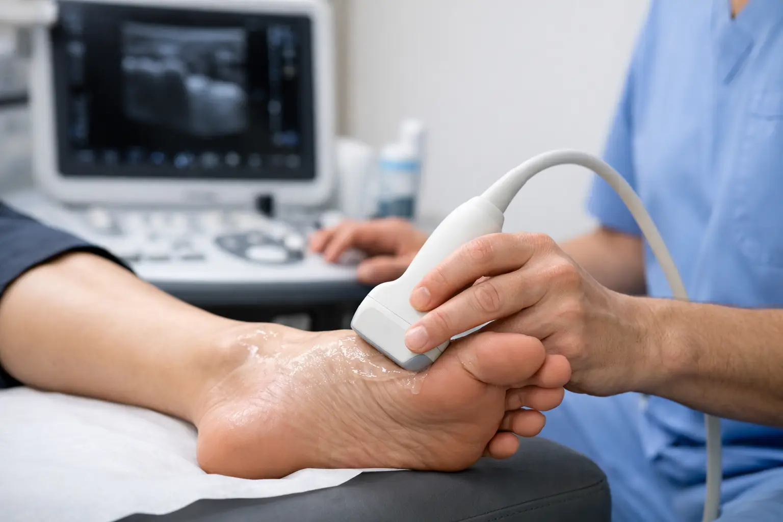
If ultrasound is indicated, gel is applied to the skin and a handheld probe is moved over the area. The scan is generally quick and well tolerated. Because it is done in real time, the clinician can explain what is being seen as the scan progresses. Patients often find this reassuring. Instead of being told that the pain is “probably” one thing or another, they can see whether a tendon is thickened, whether the plantar fascia is inflamed, or whether there is fluid around a structure that should not be there.
Why ultrasound can change treatment decisions
The main benefit of imaging is not the image itself. It is the quality of the treatment plan that follows. When the diagnosis is clearer, treatment can be more specific.
Take plantar heel pain as an example. If ultrasound shows thickening and degeneration within the plantar fascia, treatment may focus on load management, calf and foot rehabilitation, footwear changes, orthotic support and, in selected cases, injection options. If the fascia looks relatively normal but the fat pad appears irritated, the management approach may be quite different. The same symptom location does not always mean the same pathology.
Similarly, tendon pain can range from mild reactive change to more established degeneration or tearing. Those differences matter. A tendon that is overloaded but structurally intact may respond well to a graded rehabilitation programme. A tendon with more significant damage may need a more cautious plan, different expectations around recovery and closer review.
Ultrasound can also help when considering procedures. If an injection is clinically appropriate, imaging helps confirm that the target area matches the diagnosis. That improves precision and supports safer decision-making. It also makes the discussion with the patient more straightforward, because the reasoning behind treatment is visible and specific.
What ultrasound does not do
Although diagnostic ultrasound for foot pain is highly useful, it is not the right tool for everything. It does not replace a full clinical assessment, and it is not always the best scan for bone injuries, deep joint pathology or more complex cases where MRI or other investigations may be more suitable.
There are also situations where imaging can show tissue changes that are present but not necessarily causing symptoms. This is why scan findings should never be interpreted in isolation. A thickened tendon on ultrasound only becomes meaningful when matched to the patient’s pain, movement findings and history.
That balance matters. Good care is not about scanning everyone. It is about using the right investigation at the right point, then explaining clearly what the findings do and do not mean.
Common conditions ultrasound may help identify
Plantar fasciitis and plantar heel pain
Ultrasound can show plantar fascia thickening, local inflammation and tissue changes near the heel attachment. This can help distinguish classic plantar fasciitis from other causes of heel pain.
Achilles and ankle tendon problems
Pain at the back or sides of the ankle is often tendon-related, but the specific tendon and severity are not always obvious without imaging. Ultrasound can assess thickening, tears, sheath inflammation and tendon movement.
Morton’s neuroma and forefoot pain
Burning pain, tingling or the feeling of standing on a pebble may suggest nerve irritation between the toes. Ultrasound can help identify a neuroma and rule in or rule out other soft tissue causes of forefoot pain.
Bursitis, cysts and soft tissue swelling
When a lump or area of swelling is present, ultrasound helps determine whether it is fluid-filled, inflamed or associated with an underlying structure such as a tendon or joint.
A more efficient route to answers
One reason patients seek specialist assessment is speed of clarity. If you are struggling to stay active, missing runs, changing the way you walk or working around persistent pain, waiting weeks or months without a clear diagnosis can be draining. A consultation that combines clinical assessment with ultrasound when needed can shorten that uncertainty.
This does not mean every condition is solved in one visit. Some problems still require staged treatment, rehabilitation and follow-up. What changes is the quality of the starting point. Instead of trialling generic treatment and hoping it fits, the plan is built around the most likely diagnosis from the outset.
That can be especially valuable for active adults, people training for events, those returning from injury and anyone who has already been through a cycle of short-term relief followed by relapse. Clear diagnosis does not guarantee instant recovery, but it usually leads to better decisions.
Is diagnostic ultrasound for foot pain right for you?
If your symptoms are persistent, difficult to pin down, or not improving as expected, ultrasound may be worth considering as part of a specialist assessment. It is often most helpful when the diagnosis is uncertain, when more than one structure may be involved, or when treatment options depend on understanding the tissue changes more precisely.
Equally, if your condition is straightforward on examination, you may not need imaging at all. A careful clinician should be honest about that. The aim is not to sell a scan. The aim is to establish what is causing the pain and decide on the most appropriate treatment path.
For patients, that usually comes down to a simple question: do you want to keep guessing, or do you want a clearer explanation of what is driving the problem? When assessment and imaging are used properly together, the answer is often much easier to find.
Pain in the foot or ankle can affect far more than exercise. It can alter how you work, walk and cope day to day. Getting a proper explanation of the cause is often the point where recovery starts to feel more manageable.