


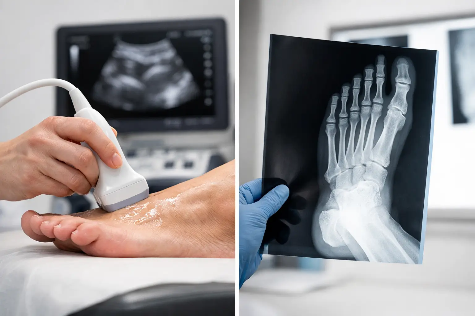
Ultrasound Scan vs Xray Foot: Which Helps?
A painful foot can look deceptively simple until you try to work out what is actually causing it. When patients search for ultrasound scan vs xray foot, they are usually asking a practical question: which test will give the clearest answer and help treatment start sooner?
The short answer is that neither scan is universally better. They do different jobs. An X-ray is best for looking at bone. Ultrasound is best for looking at soft tissue structures such as tendons, ligaments, fascia, bursae and some joints in motion. The right choice depends on your symptoms, how the injury happened, how long the problem has been there, and what a clinical examination suggests.
That is why imaging works best when it follows a proper assessment rather than being treated as a standalone purchase. A scan is most useful when it answers a specific clinical question.
Ultrasound scan vs xray foot: the core difference
If your clinician suspects a fracture, significant arthritis, a bone deformity or changes in joint alignment, an X-ray is often the more appropriate first investigation. It shows the structure of bone clearly and can reveal breaks, stress injuries that are visible on X-ray, joint degeneration, heel spurs and certain structural changes.
If the concern is plantar fasciitis, Achilles tendon pain, tendon tears, ligament injury, bursitis, Morton’s neuroma or inflammation around joints and soft tissues, ultrasound is often more informative. It shows soft tissue detail in real time and can assess structures while the foot moves or while pressure is applied to the painful area.
This matters because many common foot and ankle problems are not primarily bony problems. A patient with heel pain may assume they need an X-ray, but if the pain is coming from the plantar fascia, the more useful investigation may be ultrasound. Equally, a patient with pain after a twist or direct impact may feel they need a soft tissue scan, but an X-ray may be the sensible first step if a fracture is possible.
What an X-ray is good at
X-rays remain an important part of foot diagnosis. They are widely used because they are quick, familiar and very good at showing bone. If someone has had trauma, cannot weight bear properly, or has focal tenderness over a bone, an X-ray may be necessary to rule out a fracture or identify a bony injury.
They are also useful in longer-term problems. If a joint is stiff, enlarged or painful, an X-ray may help show arthritic change, narrowing of joint spaces, bone spurs or alignment issues. In some cases, this affects treatment planning directly. Orthotic prescription, offloading strategies and referral decisions can all change depending on what the bony architecture looks like.
The limitation is that X-rays do not show soft tissue pathology well. They cannot reliably tell you whether a tendon is thickened, whether a ligament is torn, or whether the plantar fascia is inflamed. They may be part of the picture, but they do not answer every question.
What ultrasound is good at
Ultrasound is particularly valuable in foot and ankle care because many painful conditions involve soft tissue. It can show tendon thickening, partial tears, fluid around tendons, plantar fascia changes, bursitis, neuromas and ligament injury. It can also help distinguish between conditions that feel similar to the patient but need different treatment.
For example, pain at the back of the heel might come from the Achilles tendon itself, the bursa nearby, or irritation at the tendon insertion. Pain under the heel could be plantar fasciopathy, a fascial tear, fat pad irritation or another soft tissue problem. These are not small differences. Treatment that is appropriate for one condition may be ineffective, or occasionally aggravating, for another.
One of the key advantages of ultrasound is that it is dynamic. The clinician can scan while moving the foot, compare one side to the other, and correlate the image directly with the exact point of pain. In the right clinical setting, that can make the diagnosis much clearer during the appointment itself.
Ultrasound scan vs xray foot in common symptoms
Heel pain is one of the best examples of why this comparison matters. If the pain has developed gradually, is worst with first steps, and the examination points to plantar fasciopathy, ultrasound may be more useful than an X-ray. If there has been a sudden injury or there is concern about a bony cause, then X-ray may need to come first.
Midfoot pain can be more complex. If there is swelling after injury, bruising or concern about a fracture, X-ray is usually important. If the pain is more related to tendon overload or soft tissue irritation, ultrasound may add more value.
Forefoot pain also depends on the pattern. A suspected neuroma or soft tissue inflammation between the toes may be well assessed with ultrasound. A suspected stress fracture or bony deformity may point towards X-ray. Sometimes one test gives enough information. Sometimes both have a role.
Pain around the ankle follows the same principle. Ligament sprains, tendon pathology and synovitis are often assessed very effectively with ultrasound. Bony injury after trauma may require X-ray first.
Why the best choice depends on the assessment
Patients often want the most advanced scan, but the most useful scan is the one matched to the likely diagnosis. Good imaging starts with history taking and examination. Where is the pain? What triggers it? Was there an injury? Is there swelling, instability, numbness or morning stiffness? Has it been present for days, months or years?
These details matter because imaging is not just about finding something abnormal. It is about finding the abnormality that actually explains your symptoms. Many scans show incidental changes, especially in active adults or people with long-standing wear and tear. A meaningful diagnosis comes from matching scan findings to the clinical picture.
That is why a consultation-led model is more reliable than choosing a scan in isolation. At South London Foot Clinic, diagnostic ultrasound is used when clinically indicated as part of a structured assessment, so the scan sits within a wider process of examining, explaining and planning treatment.
When you might need one, the other, or both
There are times when the choice is straightforward. A clear traumatic injury with bony tenderness may need X-ray. Ongoing tendon pain or suspected plantar fascia pathology may suit ultrasound. But foot problems are not always neat.
A patient with persistent heel pain may have soft tissue findings on ultrasound and also mechanical factors or bony changes that influence treatment. Someone with forefoot pain may need X-ray to assess structure and ultrasound to assess a neuroma or inflamed soft tissue. A runner with a suspected stress injury may need further imaging beyond either of these if symptoms and examination point that way.
This is where trade-offs matter. Ultrasound gives excellent soft tissue detail, but it does not replace X-ray for fractures and bony alignment. X-ray is strong for bone, but it can miss the soft tissue driver of pain. The right pathway is not about choosing a winner. It is about choosing the investigation that answers the next important question.
Does ultrasound replace X-ray for foot pain?
Usually, no. It complements it. In some cases, ultrasound can be the best first investigation and may remove the need for other imaging. In other cases, X-ray is essential. The decision should come from the likely diagnosis, not from a general assumption that one test is newer or better.
It is also worth remembering that more imaging is not automatically better care. If a thorough examination already makes the diagnosis clear, treatment may be able to start without any scan at all. Equally, if the presentation is uncertain, imaging can save time by narrowing down the cause and helping avoid trial-and-error treatment.
What patients should take from this
If you are comparing ultrasound scan vs xray foot, the most useful question is not which scan is best in general. It is which scan is best for your symptoms. Bone pain, fracture suspicion and structural joint issues often point towards X-ray. Tendon, fascia, ligament and other soft tissue concerns often point towards ultrasound.
The strongest diagnostic pathway is one that begins with a detailed consultation, uses imaging when it adds genuine value, and then links the findings directly to treatment. That might mean rehabilitation, orthotic support, activity modification, injection therapy, footwear advice or another targeted intervention based on the diagnosis.
When foot pain has been dragging on, uncertainty is often the hardest part. The right assessment should reduce that uncertainty, explain the cause clearly and give you a realistic plan for what happens next.